Neuroimaging
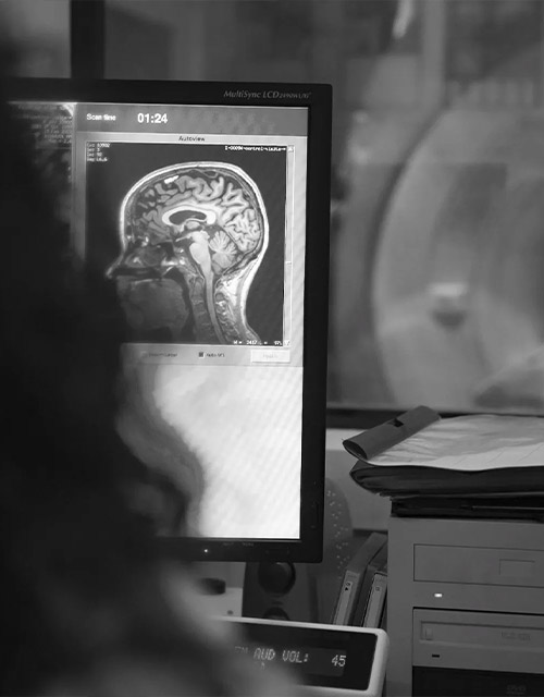
The research projects of the Neuroimaging Unit focus on advancing research on neurodegenerative diseases through the use of state-of-the-art neuroimaging techniques. Neuroimaging plays a pivotal role in unraveling the complex brain changes underlying cognitive decline and dementia. By enabling the in-vivo assessment of disease-specific patterns of brain degeneration, neuroimaging serves as an essential tool for early diagnosis, disease monitoring, and the development of personalized treatment approaches in Alzheimer’s disease and related neurodegenerative dementias.
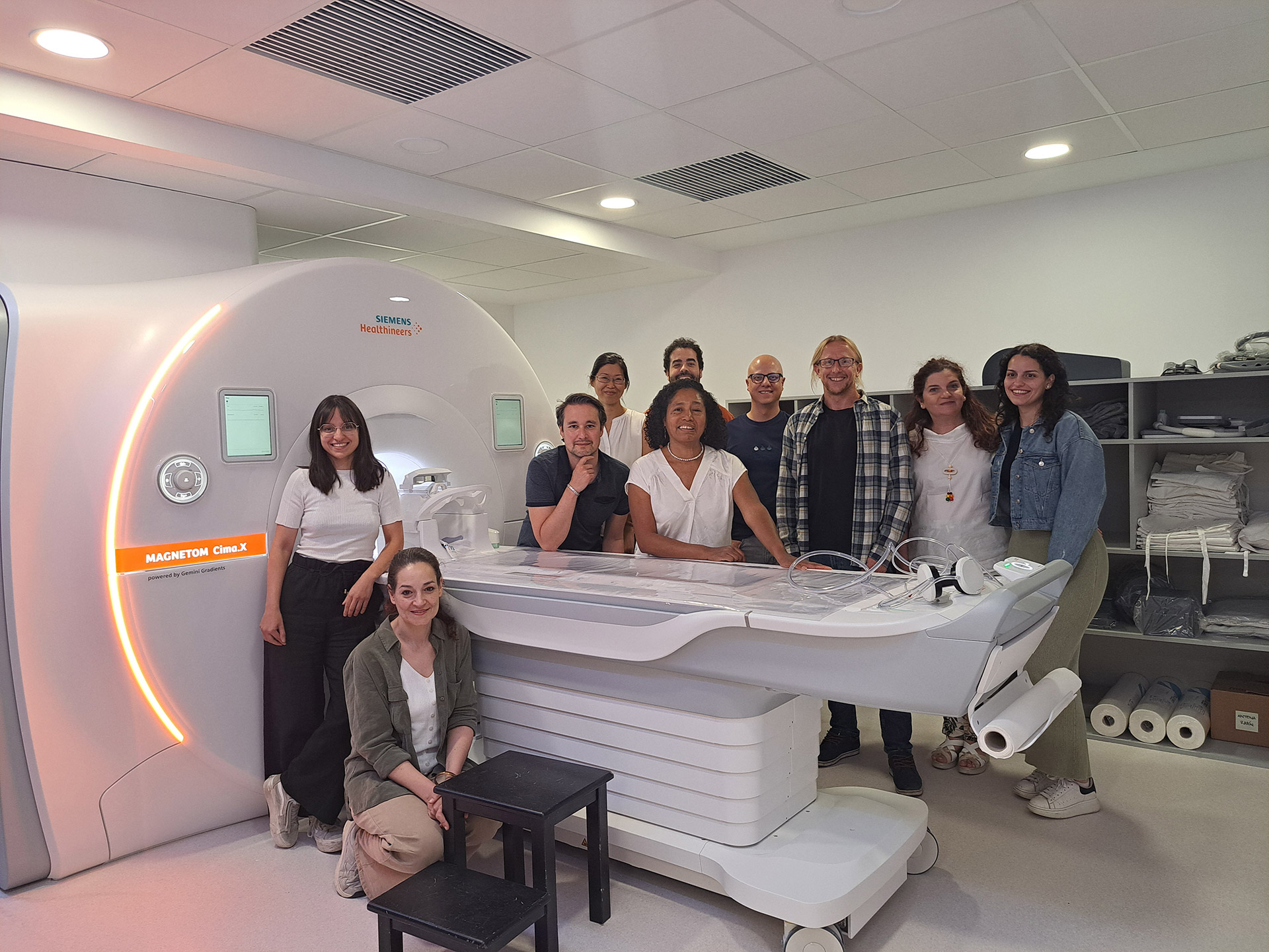
The Unit is equipped with cutting-edge infrastructure, including a Siemens Cima.X 3T MRI scanner, the first of its kind installed in a European research center. This high-performance system enables acquisition of advanced multimodal MRI sequences with exceptional resolution and speed. As the central neuroimaging core, the Unit oversees and supports all neuroimaging-related aspects of CIEN’s comprehensive research projects and cohort studies of patients with different types of neurodegenerative diseases. As such, the research activity in the Neuroimaging Unit includes coordinated acquisition, storage, pre-processing, and computational analysis of diverse types of neuroimaging data, including multimodal MRI acquisitions at the Unit’s own research-dedicated Cima.X 3T-MRI scanner as well as external acquisitions of PET imaging data at collaborating Nuclear Medicine Departments.
To carry out clinical-translational research projects, the Neuroimaging Unit has access to the rich biological samples and phenotyping data that are being collected through CIEN’s large-scale observational cohort studies of different patient populations, including the recently completed Vallecas project, which is a unique longitudinal cohort study of over 1000 deeply phenotyped older individuals who had normal cognition at baseline and underwent longitudinal clinical, multimodal MRI, and blood measurements for up to 10 years. In addition, the Vallecas Alzheimer Reina Sofia (VARS) study is a world-wide unique cohort study that follows dementia patients living in the associated nursing home of the Reina Sofia Foundation Alzheimer Centre with repeated clinical assessments, multimodal MRI, and biological sample collections, and provides detailed neuropathological examination after death in the context of a brain donation program. This design facilitates minimal time between ante-mortem neuroimaging assessments and brain collection and provides a unique opportunity to carry out neuropathological validation studies of multimodal MRI biomarkers in a cohort of dementia patients with diverse etiologies. Other ongoing cohort studies at the CIEN Foundation include the Madrid Frontotemporal Dementia Consortium and the large-scale national precision medicine project SCAP-AD, which recruits elderly individuals with early cognitive deficits that undergo in-depth neurological and neuropsychological examinations, CSF and blood sample collection, as well as a multimodal MRI-PET neuroimaging protocol.


The Neuroimaging Platform team, led by Dr Michel Grothe (PhD in Clinical Neuroscience), has a multidisciplinary character and is composed of the following professionals:
PhD in Clinical Neuroscience. PI and team leader.
PhD in Clinical Research in Medicine. Degree in Physics, expert in Neuroimaging. | Post-doctoral researcher
PhD in Radiological Imaging. Degree in Psychology. Post-doctoral researcher.
Graduate in Psychology, specialist in Neurosciences | Pre-doctoral Researcher
Graduate in Psychology, specialising in Neurosciences | Pre-doctoral Researcher
PhD in Neurosciences | Graduate in Psychology | Post-doctoral researcher
Graduate in Psychology, specialising in Neurosciences | Pre-doctoral Researcher
Coordinator. Radiodiagnostic Technician. Graduate in Radiology.
Senior technician in Diagnostic Imaging
Senior Diagnostic Imaging Technician. Graduate in Psychology.
Higher University Technician in Radiology and Imaging.
Higher Technician in Diagnostic Imaging
Neuroimaging Platform Secretary
Principal Research Lines
Our research focuses on the use of multimodal neuroimaging techniques to study the neuronal correlates and molecular underpinnings of cognitive decline and dementia in age-related neurodegenerative diseases, most notably Alzheimer’s disease (AD) and related neurodegenerative dementias. The overarching goal of this research is to better understand the diverse pathophysiologic processes that lead to cognitive decline and dementia in the elderly, and to use this information for the development of clinically useful tools for earlier and more precise differential diagnosis of age-related neurodegenerative diseases and personalized prognosis of an individual’s risk for cognitive decline and dementia.
Current core research lines at the Neuroimaging Unit include:
- Pathology-specific neuroimaging biomarkers: Imaging-pathology correlation studies for the identification of distinct multimodal neuroimaging signatures associated with Alzheimer’s disease and other neurodegenerative pathologies, including Lewy body disease, limbic age-related TDP-43 encephalopathy (LATE), and primary age-related tauopathy (PART).
- Clinical utility of in-vivo neuroimaging assessments: Different types of multimodal neuroimaging biomarkers are evaluated for their diagnostic and prognostic utility in large-scale clinical cohort studies of older individuals with incipient memory patients.
- Integration with molecular biomarkers: Combining neuroimaging features with fluid-based molecular biomarkers and genetic information to gain a better understanding of the underlying neurobiological mechanisms and to improve the precision of differential dementia diagnosis and the personalized prognosis of cognitive decline.
-
View publication
Silva-Rodríguez J, Labrador-Espinosa MA, Zhang L, Castro-Labrador S, López-González FJ, Moscoso A, Sánchez-Juan P, Schöll M, Grothe MJ.
The effect of Lewy body (co-)pathology on the clinical and imaging phenotype of amnestic patients.
Brain. 2025 Jan 31:awaf037. doi:10.1093/brain/awaf037. PMID: 39888600. Analyzes the impact of co-existing Lewy body pathology in amnestic patients, revealing differences in clinical presentation and neuroimaging profiles. -
View publication
Ortega-Cruz D, Rabano A, Strange BA.
Neuropathological contributions to grey matter atrophy and white matter hyperintensities in amnestic dementia.
Alzheimers Res Ther. 2025 Jan 9;17(1):16. doi:10.1186/s13195-024-01633-2. PMID: 39789603 Explores how various neuropathological lesions contribute to cortical atrophy and white matter hyperintensities in amnestic dementia. -
View publication
Wolk DA, Nelson PT, Apostolova L, Arfanakis K, Boyle PA, Carlsson CM, Corriveau-Lecavalier N, Dacks P, Dickerson BC, Domoto-Reilly K, Dugger BN, Edelmayer R, Fardo DW, Grothe MJ, et al.
Clinical criteria for limbic-predominant age-related TDP-43 encephalopathy.
Alzheimers Dement. 2025 Jan;21(1):e14202. doi:10.1002/alz.14202. PMID: 39807681. Presents consensus-based clinical criteria for limbic-predominant age-related TDP-43 encephalopathy (LATE), aiming to improve diagnosis and research consistency. -
View publication
Silva-Rodríguez J, Labrador-Espinosa MA, Castro-Labrador S, Muñoz-Delgado L, Franco-Rosado P, Castellano-Guerrero AM, et al.
Imaging biomarkers of cortical neurodegeneration underlying cognitive impairment in Parkinson's disease.
Eur J Nucl Med Mol Imaging. 2025 May;52(6):2002-2014. doi:10.1007/s00259-025-07070-z. PMID: 39888421. Compares imaging modalities to identify cortical biomarkers associated with cognitive impairment in Parkinson's disease. -
View publication
Labrador-Espinosa MA, Silva-Rodriguez J, Okkels N, Muñoz-Delgado L, Horsager J, Castro-Labrador S, et al.
Cortical hypometabolism in Parkinson's disease is linked to cholinergic basal forebrain atrophy.
Mol Psychiatry. 2024 Dec 5. doi:10.1038/s41380-024-02842-9. PMID: 39639173. Links cortical hypometabolism in Parkinson’s disease to atrophy of the cholinergic basal forebrain, emphasizing its role in cognitive decline. -
View publication
Garo-Pascual M, Zhang L, Valentí-Soler M, Strange BA.
Superagers Resist Typical Age-Related White Matter Structural Changes.
J Neurosci. 2024 Jun 19;44(25):e2059232024. doi:10.1523/JNEUROSCI.2059-23.2024. PMID: 38684365. Demonstrates that superagers exhibit notable resistance to typical age-related structural changes in white matter. -
View publication
Wisse LEM, Spotorno N, Rossi M, Grothe MJ, Mammana A, Tideman P, Baiardi S, et al.
MRI Signature of α-Synuclein Pathology in Asymptomatic Stages and a Memory Clinic Population.
JAMA Neurol. 2024 Oct 1;81(10):1051-1059. doi:10.1001/jamaneurol.2024.2713. PMID: 39068668. Identifies MRI features associated with α-synuclein pathology, detectable in both asymptomatic stages and memory clinic patients. -
View publication
Okkels N, Grothe MJ, Taylor JP, Hasselbalch SG, Fedorova TD, Knudsen K, et al.
Cholinergic changes in Lewy body disease: implications for presentation, progression and subtypes.
Brain. 2024 Jul 5;147(7):2308-2324. doi:10.1093/brain/awae069. PMID: 38437860. Reviews changes in the cholinergic system across different subtypes and stages of Lewy body disease and how they influence symptomatology and disease progression. -
View publication
Ortega-Cruz D, Iglesias JE, Rabano A, Strange BA.
Hippocampal sclerosis of aging at post-mortem is evident on MRI more than a decade prior.
Alzheimers Dement. 2023 Nov;19(11):5307-5315. doi:10.1002/alz.13352. PMID: 37366342. Shows that hippocampal sclerosis of aging can be detected on MRI more than a decade before death. -
View publication
Garo-Pascual M, Gaser C, Zhang L, Tohka J, Medina M, Strange BA.
Brain structure and phenotypic profile of superagers compared with age-matched older adults: a longitudinal analysis from the Vallecas Project.
Lancet Healthy Longev. 2023 Aug;4(8):e374-e385. doi:10.1016/S2666-7568(23)00079-X. PMID: 37454673. A longitudinal study from the Vallecas Project identifying brain structural features associated with successful cognitive aging.
)